Full-angle tomographic phase microscopy of flowing quasi-spherical cells†

Abstract
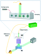
We report a reliable full-angle tomographic phase microscopy (FA-TPM) method for flowing quasi-spherical cells along microfluidic channels. This method lies in a completely passive optical system, i.e. mechanical scanning or multi-direction probing of the sample is avoided. It exploits the engineered rolling of cells while they are flowing along a microfluidic channel. Here we demonstrate significant progress with respect to the state of the art of in-flow TPM by showing a general extension to cells having almost spherical shapes while they are flowing in suspension. In fact, the adopted strategy allows the accurate retrieval of rotation angles through a theoretical model of the cells' rotation in a dynamic microfluidic flow by matching it with phase-contrast images resulting from holographic reconstructions. So far, the proposed method is the first and the only one that permits to get in-flow TPM by probing the cells with full-angle, achieving accurate 3D refractive index mapping and the simplest optical setup, simultaneously. Proof of concept experiments were performed successfully on human breast adenocarcinoma MCF-7 cells, opening the way for the full characterization of circulating tumor cells (CTCs) in the new paradigm of liquid biopsy.
 Please wait while we load your content...

