An integrated microwell array platform for cell lasing analysis†
Abstract
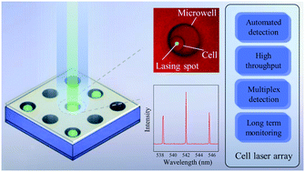
Biological cell lasers are emerging as a novel technology in biological studies and biomedical engineering. The heterogeneity of cells, however, can result in various lasing behaviors from cell to cell. Thus, the capability to track individual cells during laser investigation is highly desired. In this work, a microwell array was integrated with high-quality Fabry–Pérot cavities for addressable and automated cell laser studies. Cells were captured in the microwells and the corresponding cell lasing was achieved and analyzed using SYTO9-stained Sf9 cells as a model system. It is found that the presence of the microwells does not affect the lasing performance, but the cell lasers exhibit strong heterogeneity due to different cell sizes, cycle stages and polyploidy. Time series laser measurements were also performed automatically with the integrated microarray, which not only enables the tracking and multiplexed detection of individual cells, but also helps identify “abnormal” cells that deviate from a large normal cell population in their lasing performance. The microarrayed cell laser platform developed here could provide a powerful tool in single cell analysis using lasing emission that complements conventional fluorescence-based cell analysis.


 Please wait while we load your content...