Issue 13, 2013

A 3D microfluidic platform incorporating methacrylated gelatin hydrogels to study physiological cardiovascular cell–cell interactions

Abstract

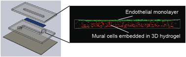
The cardiovascular system is particularly well-suited to modelling with microfluidic technologies, and much progress has been made to create microfluidic devices that mimic the microvasculature. In contrast, microfluidic platforms that model larger blood vessels and heart valves are lacking, despite the clear potential benefits of improved physiological relevance and enhanced throughput over traditional cell culture technologies. To address this need, we developed a bilayer membrane microfluidic device to model the vascular/valvular three-dimensional environment. Key features of the platform include physiologically-relevant spatial arrangement of multiple cell types, fluid flow over an endothelial monolayer, a porous membrane that permits heterotypic cell interactions while maintaining cell compartmentalization, and a photopolymerizable gelatin methacrylate (gel-MA) hydrogel as a physiologically-relevant subendothelial 3D matrix. Processing guidelines were defined for successful in-channel polymerization of gel-MA hydrogels that were mechanically stable, had physiologically-relevant elastic moduli of 2–30 kPa, and supported over 80% primary cell viability for at least four days in culture. The platform was applied to investigate shear stress-regulated paracrine interactions between valvular endothelial cells and valvular interstitial cells. The presence of endothelial cells significantly suppressed interstitial cell pathological differentiation to α-smooth muscle actin-positive myofibroblasts, an effect that was enhanced when the endothelium was exposed to flow-induced shear stress. We expect this versatile organ-on-a-chip platform to have broad utility for mechanistic vascular and valvular biology studies and to be useful for drug screening in physiologically-relevant 3D cardiovascular microenvironments.

Graphical abstract: A 3D microfluidic platform incorporating methacrylated gelatin hydrogels to study physiological cardiovascular cell–cell interactions

Article information

Article type
Paper
Submitted
10 Jan 2013
Accepted
12 Mar 2013
First published
12 Mar 2013

Lab Chip, 2013,13, 2591-2598

A 3D microfluidic platform incorporating methacrylated gelatin hydrogels to study physiological cardiovascular cell–cell interactions

M. B. Chen, S. Srigunapalan, A. R. Wheeler and C. A. Simmons, Lab Chip, 2013, 13, 2591 DOI: 10.1039/C3LC00051F

To request permission to reproduce material from this article, please go to the Copyright Clearance Center request page.

If you are an author contributing to an RSC publication, you do not need to request permission provided correct acknowledgement is given.

If you are the author of this article, you do not need to request permission to reproduce figures and diagrams provided correct acknowledgement is given. If you want to reproduce the whole article in a third-party publication (excluding your thesis/dissertation for which permission is not required) please go to the Copyright Clearance Center request page.

Read more about how to correctly acknowledge RSC content.

Social activity

Spotlight

Advertisements