Issue 21, 2012

Microfabricated mimics of in vivo structural cues for the study of guided tumor cell migration

Abstract

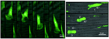
Guided cell migration plays a crucial role in tumor metastasis, which is considered to be the major cause of death in cancer patients. Such behavior is regulated in part by micro/nanoscale topographical cues present in the parenchyma or stroma in the form of fiber-like and/or conduit-like structures (e.g., white matter tracts, blood/lymphatic vessels, subpial and subperitoneal spaces). In this paper we used soft lithography micromolding to develop a tissue culture polystyrene platform with a microscale surface pattern that was able to induce guided cell motility along/through fiber-/conduit-like structures. The migratory behaviors of primary (glioma) and metastatic (lung and colon) tumors excised from the brain were monitored via time-lapse microscopy at the single cell level. All the tumor cells exhibited axially persistent cell migration, with percentages of unidirectionally motile cells of 84.0 ± 3.5%, 58.3 ± 6.8% and 69.4 ± 5.4% for the glioma, lung, and colon tumor cells, respectively. Lung tumor cells showed the highest migratory velocities (41.8 ± 4.6 μm h−1) compared to glioma (24.0 ± 1.8 μm h−1) and colon (26.7 ± 2.8 μm h−1) tumor cells. This platform could potentially be used in conjunction with other biological assays to probe the mechanisms underlying the metastatic phenotype under guided cell migration conditions, and possibly by itself as an indicator of the effectiveness of treatments that target specific tumor cell motility behaviors.

Graphical abstract: Microfabricated mimics of in vivo structural cues for the study of guided tumor cell migration

Supplementary files

Article information

Article type
Paper
Submitted
28 Jun 2012
Accepted
20 Aug 2012
First published
21 Aug 2012

Lab Chip, 2012,12, 4424-4432

Microfabricated mimics of in vivo structural cues for the study of guided tumor cell migration

D. Gallego-Perez, N. Higuita-Castro, L. Denning, J. DeJesus, K. Dahl, A. Sarkar and D. J. Hansford, Lab Chip, 2012, 12, 4424 DOI: 10.1039/C2LC40726D

To request permission to reproduce material from this article, please go to the Copyright Clearance Center request page.

If you are an author contributing to an RSC publication, you do not need to request permission provided correct acknowledgement is given.

If you are the author of this article, you do not need to request permission to reproduce figures and diagrams provided correct acknowledgement is given. If you want to reproduce the whole article in a third-party publication (excluding your thesis/dissertation for which permission is not required) please go to the Copyright Clearance Center request page.

Read more about how to correctly acknowledge RSC content.

Social activity

Spotlight

Advertisements