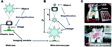
Abstract
A mini-

- This article is part of the themed collection: 2012 Lab on a Chip Emerging Investigators
Maintenance work is planned for Wednesday 1st May 2024 from 9:00am to 11:00am (BST).
During this time, the performance of our website may be affected - searches may run slowly and some pages may be temporarily unavailable. If this happens, please try refreshing your web browser or try waiting two to three minutes before trying again.
We apologise for any inconvenience this might cause and thank you for your patience.
* Corresponding authors
a
Center for Biomedical Engineering, Department of Medicine, Brigham and Women's Hospital, Harvard Medical School, Boston, MA, USA
E-mail:
alik@rics.bwh.harvard.edu
b Harvard-MIT Division of Health Sciences and Technology, Massachusetts Institute of Technology, Cambridge, MA, USA
c Wyss Institute for Biologically Inspired Engineering at Harvard University, Boston, MA, USA
d Current Address, Department of Maxillofacial Biomedical Engineering and Institute of Oral Biology, School of Dentistry, Kyung Hee University, South Korea
e Department of Mechanical Engineering, Inha University, Incheon, Republic of Korea
f Vascular Biology, Children's Hospital, Harvard Medical School, Boston, MA, 02115, USA, and Harvard School of Engineering and Applied Sciences, Cambridge, MA, USA
A mini-

 Please wait while we load your content...
Something went wrong. Try again?
S. B. Kim, K. Koo, H. Bae, M. R. Dokmeci, G. A. Hamilton, A. Bahinski, S. M. Kim, D. E. Ingber and A. Khademhosseini, Lab Chip, 2012, 12, 3976 DOI: 10.1039/C2LC40345E
To request permission to reproduce material from this article, please go to the Copyright Clearance Center request page.
If you are an author contributing to an RSC publication, you do not need to request permission provided correct acknowledgement is given.
If you are the author of this article, you do not need to request permission to reproduce figures and diagrams provided correct acknowledgement is given. If you want to reproduce the whole article in a third-party publication (excluding your thesis/dissertation for which permission is not required) please go to the Copyright Clearance Center request page.
Read more about how to correctly acknowledge RSC content.
 Fetching data from CrossRef.
This may take some time to load.
Loading related content
