Issue 13, 2011

A magnetic resonance (MR) microscopy system using a microfluidically cryo-cooled planar coil

Abstract

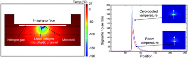
We present the development of a microfluidically cryo-cooled planar coil for magnetic resonance (MR) microscopy. Cryogenically cooling radiofrequency (RF) coils for magnetic resonance imaging (MRI) can improve the signal to noise ratio (SNR) of the experiment. Conventional cryostats typically use a vacuum gap to keep samples to be imaged, especially biological samples, at or near room temperature during cryo-cooling. This limits how close a cryo-cooled coil can be placed to the sample. At the same time, a small coil-to-sample distance significantly improves the MR imaging capability due to the limited imaging depth of planar MR microcoils. These two conflicting requirements pose challenges to the use of cryo-cooling in MR microcoils. The use of a microfluidic based cryostat for localized cryo-cooling of MR microcoils is a step towards eliminating these constraints. The system presented here consists of planar receive-only coils with integrated cryo-cooling microfluidic channels underneath, and an imaging surface on top of the planar coils separated by a thin nitrogen gas gap. Polymer microfluidic channel structures fabricated through soft lithography processes were used to flow liquid nitrogen under the coils in order to cryo-cool the planar coils to liquid nitrogen temperature (−196 °C). Two unique features of the cryo-cooling system minimize the distance between the coil and the sample: (1) the small dimension of the polymer microfluidic channel enables localized cooling of the planar coils, while minimizing thermal effects on the nearby imaging surface. (2) The imaging surface is separated from the cryo-cooled planar coil by a thin gap through which nitrogen gas flows to thermally insulate the imaging surface, keeping it above 0 °C and preventing potential damage to biological samples. The localized cooling effect was validated by simulations, bench testing, and MR imaging experiments. Using this cryo-cooled planar coil system inside a 4.7 Tesla MR system resulted in an average image SNR enhancement of 1.47 ± 0.11 times relative to similar room-temperature coils.

Graphical abstract: A magnetic resonance (MR) microscopy system using a microfluidically cryo-cooled planar coil

Supplementary files

Article information

Article type
Paper
Submitted
19 Jan 2011
Accepted
20 Apr 2011
First published
23 May 2011

Lab Chip, 2011,11, 2197-2203

A magnetic resonance (MR) microscopy system using a microfluidically cryo-cooled planar coil

C. Koo, R. F. Godley, J. Park, M. P. McDougall, S. M. Wright and A. Han, Lab Chip, 2011, 11, 2197 DOI: 10.1039/C1LC20056A

To request permission to reproduce material from this article, please go to the Copyright Clearance Center request page.

If you are an author contributing to an RSC publication, you do not need to request permission provided correct acknowledgement is given.

If you are the author of this article, you do not need to request permission to reproduce figures and diagrams provided correct acknowledgement is given. If you want to reproduce the whole article in a third-party publication (excluding your thesis/dissertation for which permission is not required) please go to the Copyright Clearance Center request page.

Read more about how to correctly acknowledge RSC content.

Social activity

Spotlight

Advertisements