High-throughput single-cell quantification using simple microwell-based cell docking and programmable time-course live-cell imaging†‡
Abstract
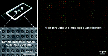
Extracting single-cell information during cellular responses to external signals in a high-throughput manner is an essential step for quantitative single-cell analyses. Here, we have developed a simple yet robust microfluidic platform for measuring time-course single-cell response on a large scale. Our method combines a simple microwell-based cell docking process inside a patterned microfluidic channel, with programmable time-course live-cell imaging and software-aided fluorescent image processing. The budding yeast, Saccharomyces cerevisiae(S. cerevisiae), cells were individually captured in microwells by multiple sweeping processes, in which a cell-containing solution plug was actively migrating back and forth several times by a finger-pressure induced receding meniscus. To optimize cell docking efficiency while minimizing unnecessary flooding in subsequent steps, circular microwells of various channel dimensions (4–24 µm diameter, 8 µm depth) along with different densities of cell solution (1.5–6.0 × 10⁹cells per mL) were tested. It was found that the microwells of 8 µm diameter and 8 µm depth allowed for an optimal docking efficiency (>90%) without notable flooding issues. For quantitative single-cell analysis, time-course (time interval 15 minute, for 2 hours) fluorescent images of the cells stimulated by mating
- This article is part of the themed collection: 10th Anniversary Issue: Korea
 Please wait while we load your content...

