Issue 24, 2009

2D-PCR: a method of mapping DNA in tissue sections

Abstract

A novel approach was developed for mapping the location of target DNA in tissue sections. The method combines a high-density, multi-well plate with an innovative single-tube procedure to directly extract, amplify, and detect the DNA in parallel while maintaining the two-dimensional (2D) architecture of the tissue. A 2D map of the gene glyceraldehyde 3-phosphate dehydrogenase (GAPDH) was created from a tissue section and shown to correlate with the spatial area of the sample. It is anticipated that this approach may be easily adapted to assess the status of multiple genes within tissue sections, yielding a molecular map that directly correlates with the histology of the sample. This will provide investigators with a new tool to interrogate the molecular heterogeneity of tissue specimens.
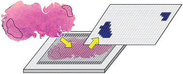
Graphical abstract: 2D-PCR: a method of mapping DNA in tissue sections

Supplementary files

Article information

Article type
Paper
Submitted
02 Jun 2009
Accepted
20 Aug 2009
First published
12 Oct 2009

Lab Chip, 2009,9, 3526-3534

2D-PCR: a method of mapping DNA in tissue sections

M. Armani, J. Rodriguez-Canales, J. Gillespie, M. Tangrea, H. Erickson, M. R. Emmert-Buck, B. Shapiro and E. Smela, Lab Chip, 2009, 9, 3526 DOI: 10.1039/B910807F

To request permission to reproduce material from this article, please go to the Copyright Clearance Center request page.

If you are an author contributing to an RSC publication, you do not need to request permission provided correct acknowledgement is given.

If you are the author of this article, you do not need to request permission to reproduce figures and diagrams provided correct acknowledgement is given. If you want to reproduce the whole article in a third-party publication (excluding your thesis/dissertation for which permission is not required) please go to the Copyright Clearance Center request page.

Read more about how to correctly acknowledge RSC content.

Social activity

Spotlight

Advertisements