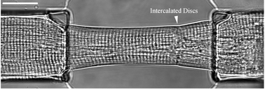
In this paper we describe a microfluidic environment that enables us to explore cell-to-cell signalling between longitudinally linked primary heart cells. We have chosen to use pairs (or doublets) of cardiac myocyte as a model system, not only because of the importance of cell–cell signalling in the study of heart disease but also because the single cardiomyocytes are both mechanically and electrically active and their synchronous activation due to the intercellular coupling within the doublet can be readily monitored on optical and electrical recordings. Such doublets have specialised intercellular contact structures in the form of the intercalated discs, comprising the adhesive junction (fascia adherens and macula adherens or desmosome) and the connecting junction (known as gap junction). The latter structure enables adjacent heart cells to share ions, second messengers and small metabolites (<1 kDa) between them and thus provides the structural basis for the synchronous (syncytical) behaviour of connected cardiomyocytes. Using the unique environment provided by the microfluidic system, described in this paper, we explore the local ionic conditions that enable the propagation of Ca2+ waves between two heart cells. We observe that the ability of intracellularCa2+ waves to traverse the intercalated discs is dependent on the relative concentrations of diastolic Ca2+ in the two adjacent cells. These experiments rely upon our ability to independently control both the electrical stimulation of each of the cells (using integrated microelectrodes) and to rapidly change (or switch) the local concentrations of ions and drugs in the extracellular buffer within the microfluidic channel (using a nanopipetting system). Using this platform, it is also possible to make simultaneous optical recordings (including fluorescence and cell contraction) to explore the effect of drugs on one or both cells, within the doublet.
You have access to this article
 Please wait while we load your content...
Something went wrong. Try again?
Please wait while we load your content...
Something went wrong. Try again?