The mouse retina in 3D: quantification of vascular growth and remodeling†
Abstract
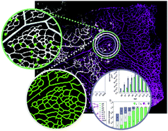
The mouse retina has become a prominent model for studying angiogenesis. The easy access and well-known developmental progression have significantly propelled our ability to examine and manipulate blood vessels in vivo. Nonetheless, most studies have restricted their evaluations to the superficial plexus (an upper vascular layer in contact with the vitreous). Here we present experimental data and quantification for the developmental progression of the full retina including the intermediate and deeper plexus that sprouts from the superficial layer. We analyze the origin and advancement of vertical sprouting and present the progression of vascular perfusion within the tissue. Furthermore, we introduce the use of Minkowsky functionals to quantify remodeling in the superficial and deeper plexus. The work expands information on the retina towards a 3D structure. This is of particular interest, as recent data have demonstrated differential effects of gene deletion on the upper and deeper plexus, highlighting the concept of distinct operational pathways during sprouting angiogenesis.
 Please wait while we load your content...