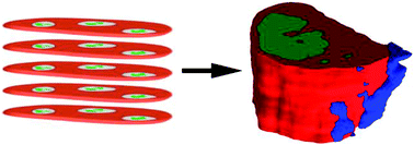
The tissue microenvironment critically influences the molecular characteristics of a tumor. However, as tumorous tissue is highly heterogeneous it may harbor various sub-populations with different microenvironments, greatly complicating the unambiguous analysis of tumor biology. Mass spectrometry imaging techniques allow for the direct analysis of tumors in the spatial context of their microenvironment. However, discovery of heterogeneous sub-populations often depends on the use of multivariate statistical methods. While this is routinely used for 2D images, multivariate statistical approaches are rarely seen in the context of 3D images. Here we present the automatic alignment of 2D images recorded by nanostructure-initiator mass spectrometry (NIMS) to reconstruct a 3D model of a mouse mammary tumor. Multivariate statistical analysis was applied to the whole 3D reconstruction at once, revealing distinct tumor regions, an observation that would not have been possible in such clarity through the analysis of isolated 2D sections. These sub-structures were confirmed by H&E and Oil Red O stains. This study shows that the combination of 3D imaging and multivariate statistics can be used to define tumor regions.
You have access to this article
 Please wait while we load your content...
Something went wrong. Try again?
Please wait while we load your content...
Something went wrong. Try again?