
Hydrothermal synthesis of core–shell structured TbPO4:Ce3+@TbPO4:Gd3+ nanocomposites for magnetic resonance and optical imaging
Abstract
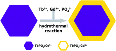
Multi-modal imaging based on multifunctional nanoparticles provides deep, non-invasive and highly sensitive imaging and is a promising alternative approach that can improve the sensitivity of early cancer diagnosis. In this study, two nanoparticles, TbPO4:Ce3+ and TbPO4:Ce3+@TbPO4:Gd3+, were synthesized via the citric-acid-mediated hydrothermal route, and then systematically characterized by means of microstructure, photoluminescence, magnetic resonance imaging (MRI), biocompatibility, and bioimaging. The results of energy dispersive X-ray spectroscopy (EDS) and electron energy loss spectroscopy (EELS) line scans indicated that TbPO4:Gd3+ nanoshells about 5 nm in thickness were successfully coated on the TbPO4:Ce3+ nanocores. X-ray diffraction (XRD) and Fourier transforms of high-resolution transmission electron microscopy (TEM) images indicated that the core–shell nanocomposites had a single crystal structure. The photoluminescence of the TbPO4:Ce3+@TbPO4:Gd3+ and TbPO4:Ce3+ nanoparticles was greatly intensified by 200 times and 100 times, respectively, compared with pure TbPO4 nanoparticles. In vitro cytotoxicity tests based on the methyl thiazolyl tetrazolium (MTT) assay demonstrated that the monodispersed nanoparticles of TbPO4:Ce3+@TbPO4:Gd3+ had low toxicity. The intracellular luminescence of the nanoparticles after being internalized by HeLa cells was also observed using confocal fluorescence microscopes. MRI showed that the nanoshells of Gd-doped TbPO4 possessed a longitudinal relaxivity of 4.067 s−1 mM−1, which is comparable to that of the commercial MRI contrast Gd-TDPA. As a result, the core–shell structured TbPO4:Ce3+@TbPO4:Gd3+ nanoparticles can potentially serve as multifunctional nanoprobes for both optical biolabels and MRI contrast agents.
 Please wait while we load your content...

