Insights from ion mobility-mass spectrometry, infrared spectroscopy, and molecular dynamics simulations on nicotinamide adenine dinucleotide structural dynamics: NAD+vs. NADH†

Abstract
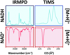
Nicotinamide adenine dinucleotide (NAD) is found in all living cells where the oxidized (NAD+) and reduced (NADH) forms play important roles in many enzymatic reactions. However, little is known about NAD+ and NADH conformational changes and kinetics as a function of the cell environment. In the present work, an analytical workflow is utilized to study NAD+ and NADH dynamics as a function of the organic content in solution using fluorescence lifetime spectroscopy and in the gas-phase using trapped ion mobility spectrometry coupled to mass spectrometry (TIMS-MS) and infrared multiple photon dissociation (IRMPD) spectroscopy. NAD solution time decay studies showed a two-component distribution, assigned to changes from a “close” to “open” conformation with the increase of the organic content. NAD gas-phase studies using nESI-TIMS-MS displayed two ion mobility bands for NAD+ protonated and sodiated species, while four and two ion mobility bands were observed for NADH protonated and sodiated species, respectively. Changes in the mobility profiles were observed for NADH as a function of the starting solution conditions and the time after desolvation, while NAD+ profiles showed no dependence. IRMPD spectroscopy of NAD+ and NADH protonated species in the 800–1800 and 3200–3700 cm−1 spectral regions showed common and signature bands between the NAD forms. Candidate structures were proposed for NAD+ and NADH kinetically trapped intermediates of the protonated and sodiated species, based on their collision cross sections and IR profiles. Results showed that NAD+ and NADH species exist in open, stack, and closed conformations and that the driving force for conformational dynamics is hydrogen bonding of the N–H–O and O–H–O forms with ribose rings.
 Please wait while we load your content...

