Structure and dysprosium dopant engineering of gadolinium oxide nanoparticles for enhanced dual-modal magnetic resonance and fluorescence imaging†
 a
a
Abstract
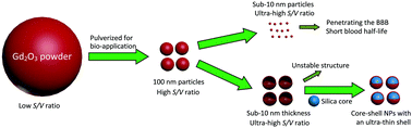
We report a class of multi-functional core–shell nanoarchitectures, consisting of silica nanospheres as the core and Gd2O3:Dy3+ nanocrystals as the ultra-thin shell, that enable unique multi-color living cell imaging and remarkable in vivo magnetic resonance imaging. These types of targeted cell imaging nanoarchitectures can be used as a variety of fluorescence nanoprobes due to the multi-color emissions of the Gd2O3:Dy3+ nanophosphor. We also proposed a strategy of modulating core–shell structure design to achieve an enhanced magnetic resonance contrast ability of Gd2O3 nanoagents, and the classical Solomon–Bloembergen–Morgan theory was applied to explicate the mechanism underlying the enhancement. The as-synthesized ligand-free nanomaterial possesses a suitable particle size for cellular uptake as well as avoiding penetrating the blood–brain barrier with good water-solubility, stability, dispersibility and uniformity. The extremely low cytotoxicity and favorable biocompatibility obtained from in vitro and in vivo bioassays of the as-designed nanoparticles indicate their excellent potential as a candidate for functioning as a targeted nanoprobe.
 Please wait while we load your content...

