
Preparation of polyacrylic acid surface-crosslinked strong fluorescent polymer nanoparticles and their sensitive in vitro imaging of cancer cells and long-life in vivo imaging of in situ tumors†
 *a
and
*a
and
Abstract
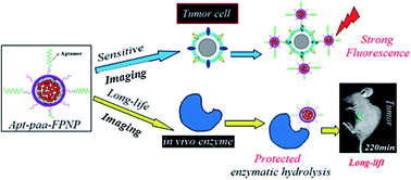
Fluorescent nanoparticles with a novel structure suitable for detecting cancer cells in vitro and in vivo have been prepared. Firstly, a strong, hydrophilic, branched amino polymer was synthesized, and then it was chemically conjugated as much as possible with fluorescein (FAM) to further synthesize a hydrophobic FAM-branched polymer with strong fluorescence. Fluorescein polymer nanoparticles (FPNPs) were then prepared based on hydrophobic interactions of the as-synthesized FAM-branched polymers, and polyacrylic acids (PAAs) were crosslinked on the surface to cover FPNPs and then to prepare PAA surface-crosslinked FPNPs (paa-FPNPs). The as-prepared paa-FPNPs nanoparticles have a fluorescent “core-cover-type” rigid structure and a non-fluorescent flexible structure consisting of the residual chains of the surface-crosslinked PAAs. The paa-FPNPs have four excellent properties as follows: (1) small particle size, a narrow particle distribution and rich negative surface charge; (2) strong fluorescence resulting from the hydrophobic interaction-based aggregation of a large number of the fluorophores conjugated on the branched amino polymers, and from a thin-cover-based weak fluorescence shielding; (3) good protection of the embedded fluorophores against in vivo enzymatic hydrolysis, owing to the outer cover and the negative residual chains of PAAs; (4) weak steric hindrance of the nanoparticle-labeled aptamer recognizing a huge-size target cell. The prepared paa-FPNPs have both a strong fluorescence and a suitable nanoparticle structure for cell imaging. Based on the ligand aptamer labeled by paa-FPNP, this paper reports the sensitive in vitro imaging of cancer cells and long-life in vivo imaging of in situ tumors.
 Please wait while we load your content...

