
Retrospective case study on the suitability of mid-infrared microscopic imaging for the diagnosis of mucormycosis in human tissue sections†
 ‡ab
‡ab
Abstract
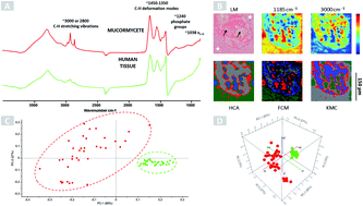
Infections caused by mucormycetes are life-threatening fungal infections, particularly for patients suffering from immunosuppression or uncontrolled diabetes mellitus. The early diagnosis of this devastating infectious fungal disease is essential to target antifungal therapy and to improve the patient's outcome. However, the diagnosis of mucormycoses remains challenging, as clinical signs and symptoms are unspecific and comprehensively evaluated diagnostic tools are missing. Therefore, we performed a retrospective case study on formalin-fixed, paraffin-embedded (FFPE) tissues of patients suffering from invasive mucormycosis, to evaluate the suitability of mid-infrared (MIR) microscopic imaging for the detection and identification of mucormycetes in human tissue sections from cheek, pleura, lung and groin by biochemical changes. 8 tissue samples of 8 patients with proven invasive mucormycosis (IM) were analysed by MIR microscopic imaging in 3 replicas. Inclusion criteria were: positive culture and/or positive molecular identification of the causative agent by the real-time PCR (polymerase chain reaction) and positive histological findings. Archived FFPE blocks of patients suffering from IM were cut and stained with Grocott. Mucormycete-positive non-stained tissue sections were chosen for MIR analysis after deparaffinization. MIR microscopic imaging is a vibrational spectroscopic technique that uses infrared radiation to image proteins and small molecules by in situ analysis in topographic maintained tissue sections. MIR microscopic imaging detects and characterizes molecules formed by the interplay of host and fungal cells. We found that MIR microscopic imaging is able to differentiate fungal elements from human tissue independent of the organ type studied. Fungal cells were detected and identified with MIR microscopic imaging.
 Please wait while we load your content...

