
Label-free imaging and identification of typical cells of acute myeloid leukaemia and myelodysplastic syndrome by Raman microspectroscopy†
Abstract
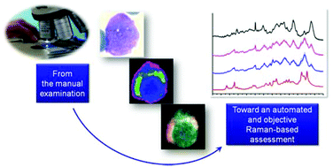
In clinical practice, the diagnosis and classification of acute myeloid leukaemia (AML) and myelodysplastic syndrome (MDS) start from the manual examination of stained smears of bone marrow (BM) and peripheral blood (PB) by using an optical microscope. This step is subjective and scarcely reproducible. Therefore, the development of subjective and potentially automatable methods for the recognition of typical AML/MDS cells is necessary. Here we have used Raman spectroscopy for distinguishing myeloblasts, promyelocytes, abnormal promyelocytes and erhytroblasts, which have to be counted for a correct diagnosis and morphological classification of AML and MDS. BM samples from patients affected by four different AML subtypes, mostly characterized by the presence of the four subpopulations selected for this study, were analyzed. First, each cell was scanned by acquiring 4096 spectra, thus obtaining Raman images which demonstrate an accurate description of morphological features characteristic of each subpopulation. Raman imaging coupled with hierarchical cluster analysis permitted the automatic discrimination and localization of the nucleus, the cytoplasm, myeloperoxidase containing granules and haemoglobin. Second, the averaged Raman fingerprint of each cell was analysed by multivariate analysis (principal component analysis and linear discriminant analysis) in order to study the typical vibrational features of each subpopulation and also for the automatic recognition of cells. The leave-one-out cross validation of a Raman-based classification model demonstrated the correct classification of myeloblasts, promyelocytes (normal/abnormal) and erhytroblasts with an accuracy of 100%. Normal and abnormal promyelocytes were distinguished with 95% accuracy. The overall classification accuracy considering the four subpopulations was 98%. This proof-of-concept study shows that Raman micro-spectroscopy could be a valid approach for developing label-free, objective and automatic methods for the morphological classification and counting of cells from AML/MDS patients, in substitution of the manual examination of BM and PB stained smears.
 Please wait while we load your content...

