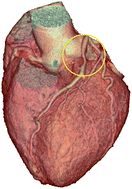
Heart disease is by far the biggest killer in the United States, and type II diabetes, which affects 8% of the U.S. population, is on the rise. In many cases, the acute coronary syndrome and/or sudden cardiac death occurs without warning. Atherosclerosis has known behavioral, genetic and dietary risk factors. However, our laboratory studies with animal models and human post-mortem tissue using FT-IR microspectroscopy reveal the chemical microstructure within arteries and in the arterial walls themselves. These include spectra obtained from the aortas of ApoE−/− knockout mice on sucrose and normal diets showing lipid deposition in the former case. Also pre-aneurysm chemical images of knockout mouse aorta walls, and spectra of plaque excised from a living human patient are shown for comparison. In keeping with the theme of the SPEC 2008 conference ‘Spectroscopic Diagnosis of Disease…’ this paper describes the background and potential value of a new catheter-based system to provide in vivo biochemical analysis of plaque in human coronary arteries. We report the following: (1) results of FT-IR microspectroscopy on animal models of vascular disease to illustrate the localized chemical distinctions between pathological and normal tissue, (2) current diagnostic techniques used for risk assessment of patients with potential unstable coronary syndromes, and (3) the advantages and limitations of each of these techniques illustrated with patent care histories, related in the first person, by the physician coauthors. Note that the physician comments clarify the contribution of each diagnostic technique to imminent cardiac risk assessment in a clinical setting, leading to the appreciation of what localized intravascular chemical analysis can contribute as an add-on diagnostic tool. The quality of medical imaging has improved dramatically since the turn of the century. Among clinical non-invasive diagnostic tools, laboratory tests of body fluids, EKG, and physical examination are still the first line of defense. However, with the fidelity of 64-slice CT imaging, this technique has recently become an option when the patient presents with symptoms of reduced arterial flow. Single photon emission computerized tomography (SPECT) treadmill exercise testing is a standard non-invasive test for decreased perfusion of heart muscle, but is time consuming and not suited for emergent evaluation. Once the invasive clinical option of catherization is chosen, this provides the opportunity for intravascular ultrasound (IVUS) imaging. As the probe is pulled through the artery, the diameter at different parts is measurable, and monochrome contrast in the constricted area reveals the presence of tissue with a different ultrasonic response. Also, via an optical catheter with a fiber-optic conductor, the possibly of spectroscopic analysis of arterial walls is now a reality. In this case, the optical transducer is coupled to a near-infrared spectrometer. Revealing the arterial chemical health means that plaque vulnerability and imminent risk could be assessed by the physician. The classical emergency use of catherization involves a contrast agent and dynamic X-ray imaging to locate the constriction, determine its severity, and possibly perform angioplasty, and stent placement.
 Please wait while we load your content...

