
Excitation modalities for enhanced micro and nanoparticle imaging in a smartphone coupled 3D printed fluorescent microscope†

Abstract
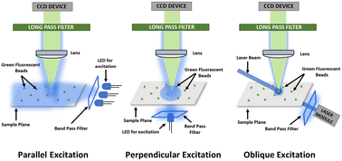
Smartphone fluorescent microscopes (SFM) offer many functional characteristics similar to their benchtop counterparts at a fraction of the cost and have been shown to work for biomarker detection in many biomedical applications. However, imaging and quantification of bioparticles in the sub-micron and nanometer range remains challenging as it requires aggressive robustness and high-performance metrics of the building blocks of SFM. Here, we explored multiple excitation modalities and their performance on the imaging capability of an SFM. Employing spatial positional variations of the excitation source with respect to the imaging sample plane (i.e., parallel, perpendicular, oblique), we developed three distinct SFM variants. These SFM variants were tested using green-fluorescent beads of four different sizes (8.3, 2, 1, 0.8 μm). Optimal excitation voltage range was determined by imaging these beads at multiple excitation voltages to optimize for no data loss and acceptable noise levels for each SFM variant. The SFM with parallel excitation was able to only image 8.3 μm beads while the SFM variants with perpendicular and oblique excitation were able to image all four bead sizes. Relative performance of the SFM variants was quantified by calculating signal difference to noise ratio (SDNR) and contrast to noise ratio (CNR) from the captured images. SFM with oblique excitation generated the highest SDNR and CNR values, whereas, for power consumption, SFM with perpendicular excitation generated the best results. This study sheds light on significant findings related to performance of SFM systems and their potential utility in biomedical applications involving sub-micron imaging. Similarly, findings of this study are translatable to benchtop microscopy instruments as well as to enhance their imaging performance metrics.
 Please wait while we load your content...

