
In vitro crystallization of calcium carbonate mediated by proteins extracted from P. placenta shells†

Abstract
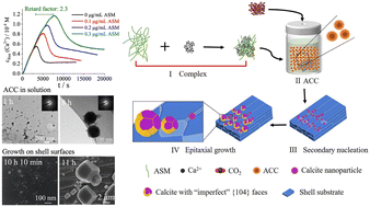
The shells of P. placenta are composed of foliated calcite laths packed as layered structures and have high mechanical properties and optical transparency. However, the biomineralization mechanism and the influence of the organic matrix on the formation of the foliated calcite laths in the shells of P. placenta are not known. The acid-soluble matrix (ASM) was extracted from the shells of P. placenta and characterized in detail. Sixteen proteins including calcium-binding and chitin-binding proteins were associated to shell formation via proteome analysis of ASM. The ASM was found to be enriched with Gly, Asp and Ser. By tracking the Ca2+ concentration in the solution in the presence of ASM by a potentiometric method, we find that the ASM has a strong effect to stabilize the amorphous calcium carbonate (ACC) precursor in solution with a nucleation time of about 7733 s and a retardation factor of about 2.3, while the concentration of the ASM was 0.3 μg mL−1. In particular, the ASM can inhibit the (secondary) nucleation of calcium carbonate for 10 and 24 hours on the shell surfaces of P. placenta and the glass substrate in the aqueous solution, respectively. An explosive secondary nucleation and quick crystal growth from 50 nm to 10 μm can be finished on the shell surfaces in one hour, in between 10 h and 11 h of reaction time, via a gas diffusion process. This indicates that the ASM has a very strong effect to stabilize ACC and inhibit nucleation. The formation of ACC in the solution and its transformation to calcite crystals on the substrates were tracked by dynamic light scattering, selected-area electron diffraction, infrared spectroscopy, scanning electron microscopy, and confocal laser scanning microscopy. It was found that the ASM was doped in the preliminary nanocrystals with a size of about 50–100 nm, which were formed on the shell surfaces via secondary nucleation, and was occluded into the calcite crystals without preferred adsorption on particular crystal planes. The calcite crystals formed on the shell surfaces became more and more rounded and lost {104} planes with increasing concentration of the ASM in the solution, indicating that the ASM can stabilize other planes instead of the thermodynamic {104} planes. The in vitro crystallization study in this work shows that ASM extracted from the shells of P. placenta has a strong effect on the biomineralization process of foliated calcite laths of P. placenta.
- This article is part of the themed collection: Biomolecular crystal engineering
 Please wait while we load your content...

