
Live-cell fluorescence imaging: assessment of thioflavin T uptake in human epidermal carcinoma cells
 a
Vijay
Sharma
*abcd
a
Vijay
Sharma
*abcd
Abstract
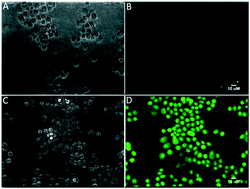
Thioflavin T (ThT), a positively charged heterocyclic small molecule, is a widely used fluorescent marker of amyloid pathophysiology to confirm the cause of death in post mortem brain tissue of Alzheimer's disease (AD) patients. Literature precedents indicate that current positron emission tomography (PET) agents, such as 11C-PIB and 18F-flutemetamol, share significant structural similarity with ThT, a lipophilic dye which does not traverse the blood–brain barrier (BBB) to enable the detection of Aβ plaques in vivo. While vital for maintaining normal physiology and healthy brain function, the BBB comprises brain endothelial cells sealed via paracellular protein complexes, bound by an extracellular matrix forming tight junctions thus controlling the delivery of molecules into the brain. The human P-glycoprotein (Pgp/ABCB1, 170 kD plasma membrane protein), belonging to the ABC family of efflux transporter proteins, also lines the luminal surface of brain endothelial cells thus poised to secrete its recognized substrates into the blood. Herein, we postulate that thioflavin T (ThT), due to its physico-chemical attributes, such as moderate lipophilicity and protonated nitrogen, could very well be recognized as a transport substrate of Pgp (P-glycoprotein, ABCB1) thus restricting its permeation into the brain. To evaluate whether or not ThT is indeed recognized by Pgp as its transport substrate thus limiting its BBB permeability, herein, we evaluate cellular accumulation profiles of ThT and PiB (a similar structural uncharged mimetic) in human epidermal carcinoma KB-3-1 (Pgp−) and MDR KB-8-5 (Pgp+) cells, using live-cell fluorescence imaging. While ThT penetrates KB-3-1 cells, it gets excluded from KB-8-5 cells, and also indicates LY335979-induced uptake in Pgp-expressing cells. Furthermore, the cellular uptake profiles of PiB are not impacted by the expression of Pgp under identical conditions. These data show that uptake profiles of ThT have been modified by the expression of Pgp in these cells, and are inversely proportional to the expression of the transporter protein located on the plasma membrane of these cells. Combined data demonstrate that ThT is efficiently recognized by Pgp as its transport substrate.
 Please wait while we load your content...