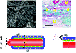
Macrophage infiltration of electrospun polyester fibers
Abstract
Electrospun fibrous polylactide (PLA) membranes have been widely used for peritendinous anti-adhesion, but their degradation may lead to granuloma formation triggered by macrophages as the result of a foreign body reaction. To understand and solve this problem in peritendinous anti-adhesion, the effects of electrospun fibrous PLA membranes (PLA-M) and the mechanism by which macrophages elicit a foreign body reaction should be elaborated. Thus, the purpose of this study was to evaluate the ability of PLA-M and ibuprofen (IBU)-loaded fibrous PLA membranes (IBU/PLA-M) to prevent adhesion/granuloma formation around the tendon, and to understand the mechanism of macrophage infiltration. The results showed that PLA-M and IBU/PLA-M were porous and that IBU could be sustainably released from IBU/PLA-M. The adhesion and proliferation of RAW264.7 macrophages were worse on the surface of IBU/PLA-M than on PLA-M. After implantation around the tendon, IBU/PLA-M had higher anti-adhesion and tendon healing scores than PLA-M. Furthermore, PLA-M was able to promote macrophage infiltration, neovascularization, TNF-α expression and collagen III deposition to a greater extent than IBU/PLA-M. Consequently, these results show that macrophages can be triggered by PLA-M which subsequently leads to inflammation and granuloma formation, while IBU/PLA-M results in enhanced anti-inflammation and anti-adhesion effects when compared to PLA-M, by reducing macrophage infiltration.

- This article is part of the themed collection: Emerging Investigators 2017


 Please wait while we load your content...