Non-staining visualization of embryogenesis and energy metabolism in medaka fish eggs using near-infrared spectroscopy and imaging

Abstract
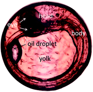
The energy metabolism and embryogenesis of fertilized Japanese medaka eggs were investigated in vivo at the molecular level using near-infrared (NIR) spectroscopy and imaging. Changes in chemical components, such as proteins and lipids, in yolk sphere and embryonic body were studied over the course of embryonic development. Metabolic changes that represent variations in the concentrations and molecular compositions of proteins and lipids in the yolk part, particularly on the 1st day after fertilization and the day just before hatching, were successfully identified in the 4900–4000 cm−1 wavenumber region. The yolk components were shown to have specific functions at the very early and final stages of the embryonic development. Proteins with α-helix- or β-sheet-rich structures clearly showed the different variation patterns within the developing egg. Furthermore, the distribution of lipids could be selectively visualized using data from the higher wavenumber region. Detailed embryonic structures were clearly depicted in the NIR images using the data from the 6400–5500 cm−1 region in which the embryo parts had some characteristic peaks due to unsaturated fatty acids. It was made clear that yolk and embryo parts had different components especially lipid components. The present study provides new insights into material variations in the fertilized egg during its growth. NIR imaging proved to be valuable in investigating the embryogenesis in vivo at the molecular level in terms of changes in biomolecular concentrations and compositions, metabolic differentiation, and detailed information about embryonic structures without the need for staining.
 Please wait while we load your content...

