
Atomic and molecular analysis highlights the biophysics of unprotonated and protonated retinal in UV and scotopic vision
Abstract
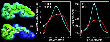
During the photoreaction of rhodopsin, retinal isomerizes, rotating the C11![[double bond, length as m-dash]](https://www.rsc.org/images/entities/char_e001.gif) C12 π-bond from cis to an all-trans configuration. Unprotonated (UR) or protonated (PR) retinal in the Schiff's base (SB) is related to UV and light vision. Because the UR and PR have important differences in their physicochemical reactivities, we compared the atomic and molecular properties of these molecules using DFT calculations. The C10–C11C12–C13 dihedral angle was rotated from 0° to 180° in 45° steps, giving five conformers, and the following were calculated from them: atomic orbital (AO) contributions to the HOMO and LUMO, atomic charges, bond length, bond order, HOMO, LUMO, hardness, electronegativity, polarizability, electrostatic potential, UV-vis spectra and dipole moment (DM). Similarly, the following were analyzed: the energy profile, hybridization, pyramidalization and the hydrogen-out-of-plane (HOOP) wagging from the H11–C11C12–H12 dihedral angle. In addition, retinal with a water H-bond (HR) in the SB was included for comparison. Interestingly, in the PR, C11 and C12 are totally the LUMO and the HOMO, respectively, and have a large electronegativity difference, which predicts an electron jump in these atoms during photoexcitation. At the same time, the PR showed a longer bond length and lower bond order, with a larger DM, lower HOMO–LUMO gap, lower hardness and higher electronegativity. In addition, the AOs of −45° and −90° conformers changed significantly, from pz to py, during the rotation concomitantly with marked hybridization, smooth pyramidalization and lower HOOP activity. Clearly, the atomic and molecular differences between the UR and PR are overwhelming, including the rotational energy profile and light absorption spectra, which indicates that light absorption of UR and PR is already determined by the retinal characteristics of the SB protonation. The HR-model compared with UR shows a lower energy barrier and a discreet bathochromic effect in the UV region.
C12 π-bond from cis to an all-trans configuration. Unprotonated (UR) or protonated (PR) retinal in the Schiff's base (SB) is related to UV and light vision. Because the UR and PR have important differences in their physicochemical reactivities, we compared the atomic and molecular properties of these molecules using DFT calculations. The C10–C11C12–C13 dihedral angle was rotated from 0° to 180° in 45° steps, giving five conformers, and the following were calculated from them: atomic orbital (AO) contributions to the HOMO and LUMO, atomic charges, bond length, bond order, HOMO, LUMO, hardness, electronegativity, polarizability, electrostatic potential, UV-vis spectra and dipole moment (DM). Similarly, the following were analyzed: the energy profile, hybridization, pyramidalization and the hydrogen-out-of-plane (HOOP) wagging from the H11–C11C12–H12 dihedral angle. In addition, retinal with a water H-bond (HR) in the SB was included for comparison. Interestingly, in the PR, C11 and C12 are totally the LUMO and the HOMO, respectively, and have a large electronegativity difference, which predicts an electron jump in these atoms during photoexcitation. At the same time, the PR showed a longer bond length and lower bond order, with a larger DM, lower HOMO–LUMO gap, lower hardness and higher electronegativity. In addition, the AOs of −45° and −90° conformers changed significantly, from pz to py, during the rotation concomitantly with marked hybridization, smooth pyramidalization and lower HOOP activity. Clearly, the atomic and molecular differences between the UR and PR are overwhelming, including the rotational energy profile and light absorption spectra, which indicates that light absorption of UR and PR is already determined by the retinal characteristics of the SB protonation. The HR-model compared with UR shows a lower energy barrier and a discreet bathochromic effect in the UV region.
 Please wait while we load your content...