Quantitative imaging of platinum based on laser ablation-inductively coupled plasma-mass spectrometry to investigate toxic side effects of cisplatin
Abstract
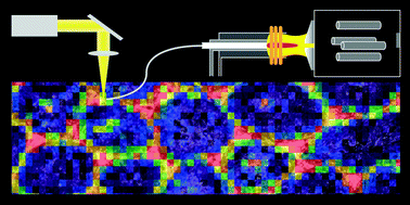
This work presents a quantitative bioimaging method for platinum based on laser ablation-inductively coupled plasma-mass spectrometry and its application for a biomedical study concerning toxic side effects of cisplatin. To trace the histopathology back to cisplatin, platinum was localized and quantified in major functional units of testicle, cochlea, kidney, nerve and brain sections from cisplatin treated mice. The direct consideration of the histology enables precise interpretation of the Pt images and the novel quantitative evaluation approach allows significantly more precise investigations than the pure image. For the first time, platinum was detected and quantified in all major injured structures including organ of Corti of cochlea and seminiferous tubule of testicle. In this way, proximal tubule in kidney, Leydig cells in testicle, stria vascularis and organ of Corti in cochlea and nerve fibers in sciatic nerves are confirmed as targets of cisplatin in these organs. However, the accumulation of platinum in almost all investigated structures also raises questions about more complex pathogenesis including direct and indirect interruption of several biological processes.
 Please wait while we load your content...