Unsupervised explorative data analysis of normal human leukocytes and BCR/ABL positive leukemic cells mid-infrared spectra†
Abstract
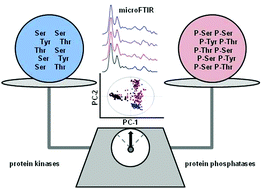
We proved the ability of Fourier Transform Infrared microspectroscopy (microFTIR) complemented by Principal Component Analysis (PCA) to detect protein phosphorylation/de-phosphorylation in mammalian cells. We analyzed by microFTIR human polymorphonuclear neutrophil (PMNs) leukocytes, mouse-derived parental Ba/F3 cells (Ba/F3#PAR), Ba/F3 cells transfected with p210BCR/ABL (Ba/F3#WT) and expressing high levels of protein tyrosine kinase (PTK), and human-derived BCR/ABL positive K562 leukemic cell sub-clones engineered to differently express receptor-type tyrosine-protein phosphatase gamma (PTPRG). Synchrotron radiation (SR) and conventional (globar) IR sources were used to perform microFTIR respectively, on single cells and over several cells within the same sample. Ex vivo time-course experiments were run, inducing maximal protein phosphorylation in PMNs by 100 nM N-formylated tripeptide fMLP. Within the specific IR fingerprint 1800–850 cm−1 frequency domain, PCA identified two regions with maximal signal variance. These were used to model and test the robustness of PCA in representing the dynamics of protein phosphorylation/de-phosphorylation processes. An IR signal ratio marker reflecting the homeostatic control by protein kinases and phosphatases was identified in normal leukocytes. The models identified by microFTIR and PCA in normal leukocytes also distinguished BCR/ABL positive Ba/F3#WT from BCR/ABL negative Ba/F3#PAR cells as well as K562 cells exposed to functionally active protein tyrosine phosphatase recombinant protein ICD-Tat transduced in cells by HIV-1 Tat technology or cells treated with the PTK inhibitor imatinib mesylate (IMA) from cells exposed to phosphatase inactive (D1028A)ICD-Tat recombinant protein and untreated control cells, respectively. The IR signal marker correctly reflected the degrees of protein phosphorylation associated with abnormal PTK activity in BCR/ABL positive leukemic cells and in general was inversely related to the expression/activity of PTPRG in leukemic sub-clones. In conclusion, we have described a new, reliable and simple spectroscopic method to study the ex vivo protein phosphorylation/de-phosphorylation balance in cell models: it is suitable for biomedical and pharmacological research labs but it also needs further optimization and its evaluation on large cohorts of patients to be proposed in the clinical setting of leukemia.
 Please wait while we load your content...

