Microfluidic device for stem cell differentiation and localized electroporation of postmitotic neurons†
Abstract
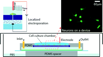
New techniques to deliver nucleic acids and other molecules for gene editing and gene expression profiling, which can be performed with minimal perturbation to cell growth or differentiation, are essential for advancing biological research. Studying cells in their natural state, with temporal control, is particularly important for primary cells that are derived by differentiation from stem cells and are adherent, e.g., neurons. Existing high-throughput transfection methods either require cells to be in suspension or are highly toxic and limited to a single transfection per experiment. Here we present a microfluidic device that couples on-chip culture of adherent cells and transfection by localized electroporation. Integrated microchannels allow long-term cell culture on the device and repeated temporal transfection. The microfluidic device was validated by first performing electroporation of HeLa and HT1080 cells, with transfection efficiencies of ~95% for propidium iodide and up to 50% for plasmids. Application to primary cells was demonstrated by on-chip differentiation of neural stem cells and transfection of postmitotic neurons with a green fluorescent protein plasmid.


 Please wait while we load your content...