
Effect of substrate choice and tissue type on tissue preparation for spectral histopathology by Raman microspectroscopy
Abstract
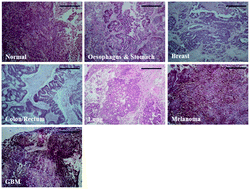
Raman spectroscopy is a non-destructive, non-invasive, rapid and economical technique which has the potential to be an excellent method for the diagnosis of cancer and understanding disease progression through retrospective studies of archived tissue samples. Historically, biobanks are generally comprised of formalin fixed paraffin preserved tissue and as a result these specimens are often used in spectroscopic research. Tissue in this state has to be dewaxed prior to Raman analysis to reduce paraffin contributions in the spectra. However, although the procedures are derived from histopathological clinical practice, the efficacy of the dewaxing procedures that are currently employed is questionable. Ineffective removal of paraffin results in corruption of the spectra and previous experiments have shown that the efficacy can depend on the dewaxing medium and processing time. The aim of this study was to investigate the influence of commonly used spectroscopic substrates (CaF2, Spectrosil quartz and low-E slides) and the influence of different histological tissue types (normal, cancerous and metastatic) on tissue preparation and to assess their use for spectral histopathology. Results show that CaF2 followed by Spectrosil contribute the least to the spectral background. However, both substrates retain paraffin after dewaxing. Low-E substrates, which exhibit the most intense spectral background, do not retain wax and resulting spectra are not affected by paraffin peaks. We also show a disparity in paraffin retention depending upon the histological identity of the tissue with abnormal tissue retaining more paraffin than normal.
 Please wait while we load your content...

