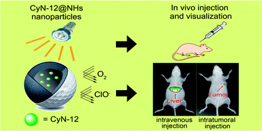
Optical near-infrared (NIR) nanomaterials provide a unique opportunity for applications in bioimaging and medical diagnosis. A kind of hydrophilic NIR fluorescent core–shell structured silica nanoparticle containing NIR cyanine chromophore, named as CyN-12@NHs, for in vivo bioimaging is developed through a facile one-pot strategy. The hydrophobic CyN-12 molecules can be successfully encapsulated into the core via the self-assembly of the amphiphilic block copolymer PS-b-PAA and subsequent shell cross-linking of silane. The as-prepared CyN-12@NHs exhibits typically spherical core–shell structure, which has a uniform size of 35 nm with a narrow size distribution, and excellent dispersity in aqueous solution. Moreover, NIR absorption (690 nm) and bright fluorescence (800 nm) of CyN-12@NHs with a large Stokes shift (110 nm) in aqueous system make it an amenable high quality bioimaging contrast agent. The core–shell nanostructure significantly enhances the chemical and photo-stability of CyN-12 via the encapsulation, which possesses a 50-times longer half-life period than free CyN-12, along with a better resistance to reactive oxygen species (ROS). Furthermore, in living cell imaging, CyN-12@NHs shows nearly no cytotoxicity and is able to outline the HepG2 cells. The in vivo imaging on a tumor-bearing mouse model indicates that CyN-12@NHs selectively accumulates in the liver after intravenous injection, and has a long retention in tumor after intra-tumor injection without decrease in fluorescence activity. Overall, the excellent photo-properties of CyN-12@NHs could meet the intricate requirements for tumor imaging, such as high sensitivity, sufficient tissue penetration, and high spatial resolution. The strategy of the silica–cyanine hybrid nanoparticles paves a desirable and efficient route to fabricate highly hydrophilic NIR fluorescent contrast agents for tumor imaging and therapy, especially with a breakthrough in photo-stability, bright fluorescence as well as large Stokes shift.


 Please wait while we load your content...