Live imaging reveals active infiltration of mitotic zone by its stem cell niche†
Abstract
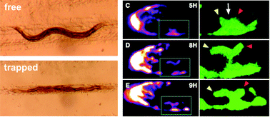
Stem cells niches are increasingly recognized as dynamic environments that play a key role in transducing signals that allow an organism to exert control on its stem cells. Live imaging of stem cell niches in their in vivo setting is thus of high interest to dissect stem cell controls. Here we report a new microfluidic design that is highly amenable to dissemination in biology laboratories that have no microfluidics expertise. This design has allowed us to perform the first time lapse imaging of the C. elegans germline stem cell niche. Our results show that this niche is strikingly dynamic, and that morphological changes that take place during development are the result of a highly active process. These results lay the foundation for future studies to dissect molecular mechanisms by which stem cell niche morphology is modulated, and by which niche morphology controls stem cell behavior.
 Please wait while we load your content...