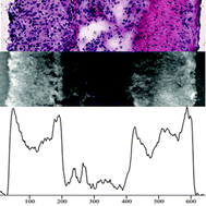
Inflammatory bowel diseases including Crohn's disease and ulcerative colitis resemble a large burden for patients due to the chronic course of disease. Therefore, there is an urgent need to explore new potential drugs and to develop new treatment options. Usually, evaluation of therapeutic potential is performed in murine models of colitis with the challenge of a valid assessment of the ongoing inflammation and the therapeutic response. Digital holographic microscopy (DHM) enables stain-free quantitative phase contrast imaging and provides tissue density assessment by measuring optical path length delay and accordingly refractive index. Dextran sodium sulphate induced colitis was performed in C57Bl/6 wildtype mice and colonic sections were examined by histological analyses and by DHM. This study proves the average refractive index to be an accurate marker to distinguish between different layers of the intestinal wall, such that the stroma is characterized by the highest value and the submucosa by the lowest. Furthermore, DHM allows a reliable detection of inflamed colonic segments (P < 0.001) with a strong correlation between the severity of inflammation and the refractive index, especially in the submucosa (R2 = 0.639). In conclusion, this approach opens a novel diagnostic option for optical quantification of inflammation in murine models of colitis. Our results pave the way to further studies to elucidate the translational potential of DHM for the clinical management of patients with inflammatory bowel diseases.
You have access to this article
 Please wait while we load your content...
Something went wrong. Try again?
Please wait while we load your content...
Something went wrong. Try again?