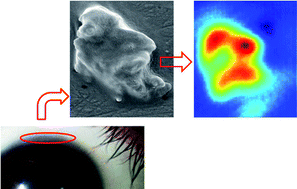
Understanding stem cell (SC) biology remains challenging and one of the few human tissues within which their in situ location is well characterized is the cornea. Individual human corneal epithelial cells were isolated from biopsies of live tissues using fluorescence-activated cell sorting (FACS); these were divided into putative SCs, transit-amplifying (TA) cells and terminally-differentiated (TD) cells. Employing synchrotron radiation-based Fourier-transform infrared (SR-FTIR) microspectroscopy with a focal plane array (FPA), sub-cellular spatial resolution analysis of unstained isolated cells was achieved as a consequence of the brilliance of a 12 collimated beams arrangement allowing rapid spectral acquisition. Infrared (IR) spectra were extracted and pre-processed. Subsequent categorization with multivariate analysis of IR spectra derived from FPA images was used to investigate biomolecular changes between classes. A progressive segregation in cell-specific spectral categories with differentiation from SC to TA cell to TD cell was noted. Multiple different absorption peaks that discriminated putative SCs, TA cells and TD cells across DNA, protein and lipid spectral regions were identified. DNA regions (1080 and 1225 cm−1) and some protein regions (1443 cm−1) primarily segregated SCs from TA cells and TD cells, whilst amide regions and lipids (1,550, 1650 and 1740 cm−1) segregated TA cells and TD cells. Scanning electron microscopy images verified the external phenotypic characteristics of the different isolated cell types. These findings highlight the applicability of SR-FTIR microspectroscopy towards distinguishing SCs, TA cells and TD cells, and suggest that cellular classification via traditional methods of immunolabelling can be greatly aided by the use of spectral biomarkers.
You have access to this article
 Please wait while we load your content...
Something went wrong. Try again?
Please wait while we load your content...
Something went wrong. Try again?


