Issue 44, 2012

Hydrodynamic deformation reveals two coupled modes/time scales of red blood cell relaxation

Abstract

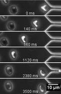
We study the mechanical relaxation behavior of human red blood cells by observing the time evolution of shape change of cells flowing through microchannels with abrupt constrictions. We observe two types of relaxation processes. In the first fast process (τ1 ∼ 200 ms) the initially parachute shaped cells relax into cup-shaped cells (stomatocytes). These cells relax and reorient in a second relaxation process with a response time of τ1/2 ∼ 10 s into the equilibrium discoid shapes. The values for the relaxation times of single red blood cells in the population scatter significantly within the cell population between 0.11 s < τ1 < 0.52 s and 9 s < τ1/2 < 49 s, respectively. However, when plotting τ1/2 against τ1, we find a linear relationship between the two timescales and are able to relate both to the elastic properties of the spectrin cytoskeleton underlying the red cell's plasma membrane. Adenosine Triphosphate (ATP) enhances dissociation of spectrin filaments resulting in a reduced shear modulus. We modify the cytoskeleton connectivity by depletion and repletion of ATP and study the effect on relaxation. Both the linear relationship of timescales as well as the ATP dependence can be understood by theoretical models.

Graphical abstract: Hydrodynamic deformation reveals two coupled modes/time scales of red blood cell relaxation

Article information

Article type
Paper
Submitted
29 Jun 2012
Accepted
17 Aug 2012
First published
12 Sep 2012

Soft Matter, 2012,8, 11240-11248

Hydrodynamic deformation reveals two coupled modes/time scales of red blood cell relaxation

S. Braunmüller, L. Schmid, E. Sackmann and T. Franke, Soft Matter, 2012, 8, 11240 DOI: 10.1039/C2SM26513C

To request permission to reproduce material from this article, please go to the Copyright Clearance Center request page.

If you are an author contributing to an RSC publication, you do not need to request permission provided correct acknowledgement is given.

If you are the author of this article, you do not need to request permission to reproduce figures and diagrams provided correct acknowledgement is given. If you want to reproduce the whole article in a third-party publication (excluding your thesis/dissertation for which permission is not required) please go to the Copyright Clearance Center request page.

Read more about how to correctly acknowledge RSC content.

Social activity

Spotlight

Advertisements