Issue 11, 2012

Comparative study of 3D morphology and functions on genetically engineered mouse melanoma cells

Abstract

Quantification of 3D morphology and measurement of cellular functions were performed on the mouse melanoma cell lines of B16F10 to investigate the intriguing problem of structure–function relations in the genetically engineered cells with GPR4 overexpression. Results of 3D analysis of cells in suspension and phase contrast imaging of adherent cells yield consistent evidence that stimulation of the proton-sensing GPR4 receptor in these cells may modify significantly their morphology with diminishing ability to produce membrane protrusions and to migrate. Examination of the 3D parameters of mitochondria provide further insights on the measured variation of the maximal capacity of oxygen consumption rate among the genetically modified cells, indicating that the proton-sensing receptor may regulate cancer cell metabolism with increased mitochondrial surface area. Our study demonstrates clearly the significant benefits of quantitative 3D morphological study in illuminating cellular functions and development of novel morphology based cell assay methods.
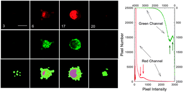
Graphical abstract: Comparative study of 3D morphology and functions on genetically engineered mouse melanoma cells

Supplementary files

Article information

Article type
Paper
Submitted
19 Jun 2012
Accepted
30 Sep 2012
First published
03 Oct 2012

Integr. Biol., 2012,4, 1428-1436

Spotlight

Advertisements