Issue 19, 2011

Polyelectrolyte multilayers generated in a microfluidic device with pH gradients direct adhesion and movement of cells

Abstract

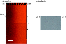
In this study, multilayers from polyethylene imine, heparin and chitosan are prepared at three different pH values of 5, 7 and 9. Water contact angle and quartz microbalance measurements show that resulting multilayers differ in terms of wetting behaviour, layer mass and mechanical properties. The multilayer is then formed within a gradient generation microfluidic (μFL) device. Polyethylene imine or heparin solutions of pH 5 are introduced into one inlet and the same solutions but at pH 9 into another inlet of the μFL device. The pH gradient established during the multilayer formation can be visualized inside the microchamber by pH sensitive fluorophores and confocal laser scanning microscopy. From this setup it is expected that properties of multilayers displayed at distinct pH values can be realised in a gradient manner inside the μFL device. Behaviour of the osteoblast cell line MG-63 seeded and cultured on top of multilayers created inside the μFL device support this hypothesis. It is observed that more cells adhere and spread on multilayers build-up at the basic side of the μFL channel, while those cells on top of multilayers built at pH 5 are fewer and smaller. These results are consistent with the behaviour of MG-63 cells seeded on multilayers formed at discrete pH values. It is particularly interesting to see that cells start to migrate from multilayers built at pH 5 to those built at pH 9 during 6 h of culture. Overall, the presented multilayer formation setup applying pH gradients leads to surfaces that promote migration of cells.

Graphical abstract: Polyelectrolyte multilayers generated in a microfluidic device with pH gradients direct adhesion and movement of cells

Supplementary files

Article information

Article type
Paper
Submitted
12 May 2011
Accepted
18 Jul 2011
First published
18 Aug 2011

Lab Chip, 2011,11, 3326-3335

Polyelectrolyte multilayers generated in a microfluidic device with pH gradients direct adhesion and movement of cells

K. Kirchhof, A. Andar, H. B. Yin, N. Gadegaard, M. O. Riehle and T. Groth, Lab Chip, 2011, 11, 3326 DOI: 10.1039/C1LC20408D

To request permission to reproduce material from this article, please go to the Copyright Clearance Center request page.

If you are an author contributing to an RSC publication, you do not need to request permission provided correct acknowledgement is given.

If you are the author of this article, you do not need to request permission to reproduce figures and diagrams provided correct acknowledgement is given. If you want to reproduce the whole article in a third-party publication (excluding your thesis/dissertation for which permission is not required) please go to the Copyright Clearance Center request page.

Read more about how to correctly acknowledge RSC content.

Social activity

Spotlight

Advertisements