Issue 4, 2011

A microfluidic system with optical laser tweezers to study mechanotransduction and focal adhesion recruitment

Abstract

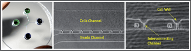
We present a new method to locally apply mechanical tensile and compressive force on single cells based on integration of a microfluidic device with an optical laser tweezers. This system can locate a single cell within customized wells exposing a square-like membrane segment to a functionalized bead. Beads are coated with extracellular matrix (ECM) proteins of interest (e.g. fibronectin) to activate specific membrane receptors (e.g. integrins). The functionalized beads are trapped and manipulated by optical tweezers to apply mechanical load on the ECM-integrin-cytoskeleton linkage. Activation of the receptor is visualized by accumulation of expressed fluorescent proteins. This platform facilitates isolation of single cells and excitation by tensile/compressive forces applied directly to the focal adhesionvia specific membrane receptors. Protein assembly or recruitment in a focal adhesion can then be monitored and identified using fluorescent imaging. This platform is used to study the recruitment of vinculin upon the application of external tensile force to single endothelial cells. Vinculin appears to be recruited above the forced bead as an elliptical cloud, centered 2.1 ± 0.5 μm from the 2 μm bead center. The mechanical stiffness of the membrane patch inferred from this measurement is 42.9 ± 6.4 pN μm−1 for a 5 μm × 5 μm membrane segment. This method provides a foundation for further studies of mechanotransduction and tensile stiffness of single cells.

Graphical abstract: A microfluidic system with optical laser tweezers to study mechanotransduction and focal adhesion recruitment

Supplementary files

Article information

Article type
Paper
Submitted
07 Oct 2010
Accepted
17 Nov 2010
First published
08 Dec 2010

Lab Chip, 2011,11, 684-694

A microfluidic system with optical laser tweezers to study mechanotransduction and focal adhesion recruitment

P. Honarmandi, H. Lee, M. J. Lang and R. D. Kamm, Lab Chip, 2011, 11, 684 DOI: 10.1039/C0LC00487A

To request permission to reproduce material from this article, please go to the Copyright Clearance Center request page.

If you are an author contributing to an RSC publication, you do not need to request permission provided correct acknowledgement is given.

If you are the author of this article, you do not need to request permission to reproduce figures and diagrams provided correct acknowledgement is given. If you want to reproduce the whole article in a third-party publication (excluding your thesis/dissertation for which permission is not required) please go to the Copyright Clearance Center request page.

Read more about how to correctly acknowledge RSC content.

Social activity

Spotlight

Advertisements