Issue 21, 2010

Characterisation and performance of hydrogel tissue scaffolds

Abstract

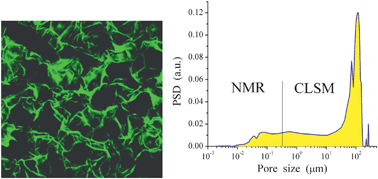
Porosity over a broad range (typically 0.001–300 μm in diameter) of tissue scaffolds provides appropriate conditions for diffusion and adsorption of small molecules and macromolecules, migration of cells through the scaffold, and adequate cell proliferative capacity. Characterisation of pores over this large range poses a problem especially when analysing soft polymer hydrogels, as no one methodology can adequately cover the entire range. This work describes a combined technique used for evaluation of the porous structure of a collagen hydrogel (dermal substitute Integra®) on the basis of NMR-cryoporometry (sensitive to nanopores) and confocal laser scanning microscopy (CLSM) imaging (sensitive to macropores). Thermodesorption of water, diffusion of proteins through a collagen membrane, migration and growth of normal primary human skin fibroblasts, and the interaction kinetics of 3T3 mouse fibroblast cells (using a quartz crystal microbalance) with collagen were analysed with respect to the porous structure of the material. The contribution to the total porosity of pores with a diameter of less than 100 nm is low, at approximately 3–5%, a figure estimated using the methods described above. However, these pores are the main contributor to the specific surface area (S ≈ 120 m2 g−1) as larger diameter macropores, with diameters of 20–200 μm, have a much lower surface area at S ≈ 0.4 m2 g−1 relative to their large pore volume V = 14.4 cm3 g−1.

Graphical abstract: Characterisation and performance of hydrogel tissue scaffolds

Article information

Article type
Paper
Submitted
02 Jul 2010
Accepted
13 Aug 2010
First published
06 Sep 2010

Soft Matter, 2010,6, 5351-5358

Characterisation and performance of hydrogel tissue scaffolds

V. M. Gun'ko, L. I. Mikhalovska, I. N. Savina, R. V. Shevchenko, S. L. James, P. E. Tomlins and S. V. Mikhalovsky, Soft Matter, 2010, 6, 5351 DOI: 10.1039/C0SM00617C

To request permission to reproduce material from this article, please go to the Copyright Clearance Center request page.

If you are an author contributing to an RSC publication, you do not need to request permission provided correct acknowledgement is given.

If you are the author of this article, you do not need to request permission to reproduce figures and diagrams provided correct acknowledgement is given. If you want to reproduce the whole article in a third-party publication (excluding your thesis/dissertation for which permission is not required) please go to the Copyright Clearance Center request page.

Read more about how to correctly acknowledge RSC content.

Social activity

Spotlight

Advertisements