Issue 2-3, 2010

Non-invasive microfluidic gap junction assay

Abstract
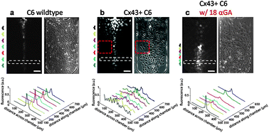
Gap junctions are protein channels between cells that allow direct electrical and metabolic coupling via the exchange of biomolecules and ions. Their expression, though ubiquitous in most mammalian cell types, is especially important for the proper functioning of cardiac and neuronal systems. Many existing methods for studying gap junction communication suffer from either unquantifiable data or difficulty of use. Here, we measure the extent of dye spread and effective diffusivities through gap junction connected cells using a quantitative microfluidic cell biology platform. After loading dye by hydrodynamic focusing of calcein/AM, dye transfer dynamics into neighboring, unexposed cells can be monitored via timelapse fluorescent microscopy. By using a selective microfluidic dye loading over a confluent layer of cells, we found that high expression of gap junctions in C6 cells transmits calcein across the monolayer with an effective diffusivity of 3.4 × 10⁻¹³ m2/s, which are highly coupled by Cx43. We also found that the gap junction blocker 18α-GA works poorly in the presence of serum even at high concentrations (50 µM); however, it is highly effective down to 2.5 µM in the absence of serum. Furthermore, when the drug is washed out, dye spread resumes rapidly within 1 min for all doses, indicating the drug does not affect transcriptional regulation of connexins in these Cx43+ cells, in contrast to previous studies. This integrated microfluidic platform enables the in situ monitoring of gap junction communication, yielding dynamic information about intercellular molecular transfer and pharmacological inhibition and recovery.

Graphical abstract: Non-invasive microfluidic gap junction assay

Article information

Article type
Technical Innovation
Submitted
21 Sep 2009
Accepted
06 Jan 2010
First published
09 Feb 2010

Integr. Biol., 2010,2, 130-138

Spotlight

Advertisements