Resonance Raman microscopy in combination with partial dark-field microscopy lights up a new path in malaria diagnostics
Bayden R.
Wood
,
Antje
Hermelink
,
Peter
Lasch
,
Keith R.
Bambery
,
Grant T.
Webster
,
Mehdi Asghari
Khiavi
,
Brian M.
Cooke
,
Samantha
Deed
,
Dieter
Naumann
and
Don
McNaughton
Analyst, 2009, 134, 1119-1125
DOI:
10.1039/B822603B
Received
16 Dec 2008,
Accepted
19 Mar 2009
First published on the web
31 Mar 2009
This article is part of the collection:
Optical Diagnosis
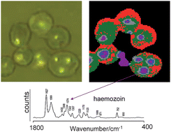
Our goal is to produce a rapid and accurate diagnostic tool for malaria using resonance Raman spectroscopy to detect small inclusions of haemozoin in Plasmodium falciparum infected red blood cells. In pursuit of this aim we serendipitously discovered a partial dark-field effect generated by our experimental setup, which helps identify in thick blood films potential parasites that are normally difficult to see with conventional bright-field microscopy. The haemozoin deposits “light up” and these can be selectively targeted with the Raman microscope to confirm the presence or absence of haemozoin by the strong 1569 cm–1 band, which is a marker for haemozoin. With newly developed imaging Raman microscopes incorporating ultra-sensitive rapid readout CCDs it is possible to obtain spectra with a good signal-to-noise ratio in 1 second. Moreover, images from a smear of potentially infected cells can be recorded and analysed with multivariate methods. The reconstructed images show what appear to be sub-micron-inclusions of haemozoin in some cells indicating that the technique has potential to identify low pigmented forms of the parasite including early trophozoite-stage infected cells. Further work is required to unambiguously confirm the presence of such forms through systematic staining but the results are indeed promising and may lead to the development of a new Raman-based malaria diagnostic.
Fetching data from CrossRef. This may take some time to load.