Micro-well arrays for 3D shape control and high resolution analysis of single cells†
Abstract
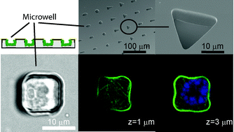
In addition to rigidity, matrix composition, and cell shape, dimensionality is now considered an important property of the cell microenvironment which directs cell behavior. However, available tools for cell culture in two-dimensional (2D) versus three-dimensional (3D) environments are difficult to compare, and no tools exist which provide 3D shape control of single cells. We developed polydimethylsiloxane (PDMS) substrates for the culture of single cells in 3D arrays which are compatible with high-resolution microscopy. Cell adhesion was limited to within microwells by passivation of the flat upper surface through ‘wet-printing’ of a non-fouling polymer and backfilling of the wells with specific adhesive proteins or lipid bilayers. Endothelial cells constrained within microwells were viable, and intracellular features could be imaged with high resolution objectives. Finally, phalloidin staining of actin stress fibers showed that the cytoskeleton of cells in microwells was 3D and not limited to the cell–substrate interface. Thus, microwells can be used to produce microenvironments for large numbers of single cells with 3D shape control and can be added to a repertoire of tools which are ever more sought after for both fundamental biological studies as well as high throughput cell screening assays.


 Please wait while we load your content...