Issue 3, 2006

Electrochemical microfluidic biosensor for the detection of nucleic acid sequences

Abstract

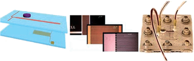
A microfluidic biosensor with electrochemical detection for the quantification of nucleic acid sequences was developed. In contrast to most microbiosensors that are based on fluorescence for signal generation, it takes advantage of the simplicity and high sensitivity provided by an amperometric and coulorimetric detection system. An interdigitated ultramicroelectrode array (IDUA) was fabricated in a glass chip and integrated directly with microchannels made of poly(dimethylsiloxane) (PDMS). The assembly was packaged into a Plexiglas® housing providing fluid and electrical connections. IDUAs were characterized amperometrically and using cyclic voltammetry with respect to static and dynamic responses for the presence of a reversible redox couple—potassium hexacyanoferrate (II)/hexacyanoferrate (III) (ferri/ferrocyanide). A combined concentration of 0.5 µM of ferro/ferricyanide was determined as lower limit of detection with a dynamic range of 5 orders of magnitude. Background signals were negligible and the IDUA responded in a highly reversible manner to the injection of various volumes and various concentrations of the electrochemical marker. For the detection of nucleic acid sequences, liposomes entrapping the electrochemical marker were tagged with a DNA probe, and superparamagnetic beads were coated with a second DNA probe. A single stranded DNA target sequence hybridized with both probes. The sandwich was captured in the microfluidic channel just upstream of the IDUA via a magnet located in the outside housing. Liposomes were lysed using a detergent and the amount of released ferro/ferricyanide was quantified while passing by the IDUA. Optimal location of the magnet with respect to the IDUA was investigated, the effect of dextran sulfate on the hybridization reaction was studied and the amount of magnetic beads used in the assay was optimized. A dose response curve using varying concentrations of target DNA molecules was carried out demonstrating a limit of detection at 1 fmol assay−1 and a dynamic range between 1 and 50 fmol. The overall assay took 6 min to complete, plus 15–20 min of pre-incubation and required only a simple potentiostat for signal recording and interpretation.

Graphical abstract: Electrochemical microfluidic biosensor for the detection of nucleic acid sequences

Article information

Article type
Paper
Submitted
19 Sep 2005
Accepted
09 Jan 2006
First published
24 Jan 2006

Lab Chip, 2006,6, 414-421

Electrochemical microfluidic biosensor for the detection of nucleic acid sequences

V. N. Goral, N. V. Zaytseva and A. J. Baeumner, Lab Chip, 2006, 6, 414 DOI: 10.1039/B513239H

To request permission to reproduce material from this article, please go to the Copyright Clearance Center request page.

If you are an author contributing to an RSC publication, you do not need to request permission provided correct acknowledgement is given.

If you are the author of this article, you do not need to request permission to reproduce figures and diagrams provided correct acknowledgement is given. If you want to reproduce the whole article in a third-party publication (excluding your thesis/dissertation for which permission is not required) please go to the Copyright Clearance Center request page.

Read more about how to correctly acknowledge RSC content.

Social activity

Spotlight

Advertisements