Bing Shi‡
a,
Di Luan‡a,
Shihui Wanga,
Lingyun Zhaob,
Lei Taoc,
Qipeng Yuana and
Xing Wang*a
aThe State Key Laboratory of Chemical Resource Engineering, Beijing University of Chemical Technology, Beijing 100029, P. R. China. E-mail: wangxing@mail.buct.edu.cn
bKey Laboratory of Advanced Materials, Ministry of Education, Institute of Regenerative Medicine and Biomimetic Material Science and Technology, Tsinghua University, Beijing 100084, P. R. China
cThe Key Laboratory of Bioorganic Phosphorus Chemistry & Chemical Biology (Ministry of Education), Department of Chemistry, Tsinghua University, Beijing 100084, P. R. China
First published on 2nd June 2015
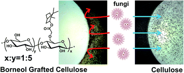
Cellulose fibers modified to obtain antifungal activity have attracted much attention due to their versatile applications, although there are still some problems associated with the conventional protocols, including the toxicity to organisms, unwanted resistance, and gradually increasing environmental pressure. A natural and safe strategy is desired to design new types of antifungal cellulose. Herein, we present a borneol-grafted cellulose (BGC) by covalently tethering L-borneol, a natural product, to cellulose. The attained BGC has been characterized to confirm the chemical and amorphous features using a combination of spectroscopic and analytical techniques. This BGC material was subsequently challenged with Mucor racemosus and Aspergillus niger, and exhibited a remarkable performance in antifungal adhesion and fungal growth inhibition, suggesting the grafted borneol moieties are crucial for influencing the tactile sensing of fungal cells and subsequent selective inadhesion. The BGC has also been evaluated as a non-cytotoxic material, implying its great potential for biomedical and sanitary applications.
Ideal antifungal materials or surfaces should be biocompatible and environment-friendly. Thus, the application of natural antimicrobials has been recognized as a suitable strategy with the popular environmental protection movement. As one of the most studied and excellent natural materials,13,14 cellulose fiber has been employed in broad fields, including textiles,15 food packing,16 medical accessory materials,17 and the environmental domain.18 Meanwhile, cellulose is also known as a good culture material for microorganisms, owning to its large surface area and moisture absorbability. For safety reasons, the antifungal issue must be considered for cellulose fibers, especially in biomedical and healthcare areas.19 Natural compositions including chitosan,20 natural dyes,21 and essential oils22 have been used to modify cellulose fibers and demonstrated promising results. However, most of the current applied natural materials still mainly depend on their poisoning effect on the cells or disrupting the integrity of cell membranes to act as antimicrobials, which hamper the application of those natural materials in broader fields.
As a broad-spectrum defensive strategy to prevent antifungal adhesion, utilizing of the “tactile sense” of fungal cells to prevent their adhesion in the initial phase has not been extensively studied. Our recent work has demonstrated that the cells exhibited a distinct inclination towards different chiral stereochemistry during their adhesion on external surfaces,23,24 which inspired us to make further efforts in developing antimicrobial materials or surfaces catering to the “chiral taste” of microbe cells. A model system using polyborneolacrylate was thus achieved,25 where an L-borneol containing polymer with good biocompatibility was proven to be a promising candidate for bacterial adhesion inhibition. As a well-known safe and common natural product, L-borneol is suitable for use based on the natural strategy for endowing antimicrobial activity to cellulose. In the current study, a borneol-grafted cellulose (BGC) has been synthesized for the first time by covalently tethering L-borneol onto the cellulose surface (Scheme 1). The antifungal activity of the modified cellulose has been systematically studied, demonstrating a remarkable performance in antifungal adhesion and fungal growth inhibition. Considering the excellent and general antimicrobial effect and that L-borneol is safe, easily available and biocompatible, this BGC composite might have potential to be used as a new generation antifungal material in the medical and clinical fields.
Scheme 1 Synthesis of chloroacetyl-L-borneol (CAB) and subsequently grafting to cellulose to obtain the borneol-grafted cellulose (BGC) via an etherification method. |
To evaluate the antifungal adhesion activity of the BGC sample, the BGC powder was pressed into 13 mm diameter sized pellets, where cellulose was used as a control for the same test. The two kinds of pellets were individually affixed onto the same beef extract peptone solid medium and then, 10 μL of fungal solution was dropped in the center of the plate, by controlling the distance to the test samples. The plate was aerobically cultured at 37 °C in an electro-heating standing-temperature cultivator. The fungi were allowed to grow and expand from the medium center to the pellet surface. Fungal growth at different periods was observed and recorded with a camera.
Scanning electron microscopy (SEM, Hitachi S-4700) was used to study the morphologies of fungal cells on the sample surface. The aforementioned samples were immobilized with 2.5% glutaraldehyde for 2 h at 4 °C. The fungal cells were dehydrated using 50, 60, 70, 80, 90 and 100% ethanol for 10 min, respectively. The samples were freeze-dried for 12 h prior to SEM observation.
The reaction occurrence was first assessed using FT-IR. Fig. 1(a) shows typical spectra of the BGC sample and the cellulose. A significant peak at 1727 cm−1, corresponding to the CO stretching of the ester, was detected in the BGC sample, indicating the grafting of CAB onto cellulose. Consequently, an increased vibration peak at 2962 cm−1, which corresponds to the C–H stretching of methyl or methylene groups was noticed in the spectral characteristics of the borneol pendants. Meanwhile, compared with the peaks of the cellulose sample, the BGC sample yielded a new peak at 1190 cm−1, corresponding to the signal of C–O–C stretching of the linking groups resulting from the etherification reaction, thus confirming the successful grafting of borneol onto cellulose. In addition, all of them were characterized with a dominant O–H stretching vibration (3070–3570 cm−1), C–H stretching vibration (2960 cm−1) and C–O stretching vibration (1029 cm−1) because of the presence of basic glucose units.
Fig. 1 (a) FT-IR spectra of BGC (top) and cellulose (bottom). (b) 13C NMR spectra of cellulose (bottom), BGC (middle) and CAB (top). |
More visual evidence was supplied by 13C NMR detection. Fig. 1(b) shows the analyzed results of the 13C NMR spectra of the final BGC product and the control groups of cellulose and CAB molecules. In comparison with the cellulose’s spectrum that is marked with band a, b, and c for the typical carbon groups, the BGC sample exhibited two remarkable signals located at δ 20 ppm (band d) and δ 173 ppm (band f), which could be attributed to methyl/methylene and ester groups, respectively. Meanwhile, the signal of the ether linkage (C–O–C) was found at δ 68.3 ppm (band e). 13C NMR detection of the CAB molecules showed three key bands located at δ 41.3, 167.5, and 80.7 ppm. The carbon near the halogen (C–Cl) contributed a band g at δ 41.3 ppm, which had disappeared on the BGC’s spectrum as expected. While, another two key bands (k and l) could be also found in the BGC’s spectrum rather than the cellulose’s spectrum, demonstrating a proper BGC structure.
The XPS spectra (Fig. 2) provided further proof for the grafting of CAB onto cellulose. For the BGC sample, the C 1s signal showed a new peak located at 283 eV that corresponds to the C–C/C–H groups of borneol. Whereas, no signal of Cl elements was found at the range of 197–202 eV,30 which was in agreement with the NMR measurements. Quantitative analysis for borneol grafting was performed using EDS measurements (Fig. S1 in ESI†), which demonstrated the elemental content of the BGC sample. It is well known that cellulose is formed from repetitive glucose units and borneol is mainly composed of carbon elements. Therefore, the grafting rate could be calculated based on the changing of the C/O element ratio. After borneol grafting, the C/O ratio in BGC (C: 52.77 wt%, O: 47.23 wt%) was increased to 1.12 compared with the ratio of 0.89 for cellulose (C: 47.13 wt%, O: 52.87 wt%). With the help of a ChemBioDraw simulation program (see Fig. S2 in ESI†), the borneol-grafting rate was calculated to be approximately 17%, in other words, every six glucose units had one CAB molecule. In addition, no signal of Cl elements was detected at 2.1269 kV.
As the cage structure of borneol was grafted onto cellulose, changes in the internal crystallinity were studied using XRD observations. Fig. S3 in the ESI† shows the full XRD patterns of the samples before and after the borneol grafting onto cellulose. The typical peaks of cellulose at 2θ = 14.5°, 16.0°, 22.5° represented the 101, 10, 002 diffraction planes respectively, corresponding to cellulose I. After borneol grafting, wide deformation was observed mainly due to two distinct reasons. Firstly, cellulose dissolved and precipitated in the ionic liquid, resulting in a crystal structure change. More importantly, borneol is a bicyclic monoterpene and this cage molecule caused the macromolecular structure to be disordered.
To investigate the adhesion details, SEM measurements were carried out to study the morphologies of M. racemosus. Fig. 4 shows the SEM images of the fungal cell adhesion on the above-mentioned pellets. A large amount of grown sporangia and hypha were found on the cellulose surface (Fig. 4a) and a lively germination of spores was exhibited therein (Fig. 4a′ and the inset). On the other hand, on the BGC surface (Fig. 4b), only a few hyphae could be observed near the boundary and the serendipitous spores stayed near the hypha presenting a whole sphericity (Fig. 4b′), indicating a growth inhibition of spores on the BGC surface. At least, these phenomena demonstrated the success of the cellulose modification and the high antifungal power of the grafted borneol molecules. It is obvious that the grafting ratio of the borneol of BGC could affect the antifungal activity. But to our surprise, the antifungal activity of BGC is achieved with a relatively low grafting rate (17%) of borneol pendants as proven in the above-mentioned analysis. As expected, if the grafting ratio of borneol decreased to approximately 6.2% (Fig. S4 in ESI†), a lesser antifungal effect could be observed (Fig. S5 in ESI†).
As mentioned, the grafting of L-borneol on cellulose was realized by employing a short acetyl linker. It is not consistent with the offensive strategy, where surface-tethered antifungal agents need to have a deep range of penetration afforded by longer anchoring linkers in order to reach the fungal cell membrane, considering the thickness of the outer protective fungal envelope (∼100 nm).2 Therefore, the interaction should happen on the outside of the fungal envelope. According to the aforementioned phenomena, we could find that the BGC mainly performs a defensive antifungal effect. Based on the understanding that cells could distinguish stereochemical signals of external surfaces,23–25 we deduced that a biological surface recognition may be a probable explanation for the antifungal action of our BGC sample. In other words, fungal cells recognized the caged bicyclic borneol molecules and subsequently tended to inadhesion on the BGC surface, where borneol grafting plays the key role in the antifungal effect of the BGC product.
In order to further demonstrate the antifungal properties, we subsequently performed the experiment with A. niger. Considering that the BGC is an antifungal pellet, the operation was carried out in ambient conditions. After 8 days of incubation, a clear surface with a distinct boundary was found for the BGC pellet (Fig. 5), while the cellulose pellet was covered with dense A. niger cells. To our surprise, other fungi such as M. racemosus and yeast that might come from the environment were found in the plate, but more importantly, the yeast cells did not tend to adhere to the BGC pellet. Through careful checking under a microscope (Fig. 5, the insets a, b and c), only a few A. niger cells were found at the boundary of the BGC pellet, but their amount was much less compared with the cellulose pellet. Based on the aforementioned mechanism, we speculated that the appearance of those cells was not due to the active adhesion of fungal cells. In any case, these phenomena distinctly exhibited a conversion of the antifungal activity from the surrender of cellulose to the resistance of BGC.
Fig. 6 MTT results of the RGR of L929 cells for 2 days of incubation in BGC conditioned media. The data are presented as the mean plus or minus the standard deviation (n = 9 for each group). |
Sample | Days of culture | Relative growth rate (%) | Toxicity grade |
---|---|---|---|
Control | 2 | 99.71 ± 2.75 | 1 |
Borneol | 2 | 98.53 ± 3.93 | 1 |
Cellulose | 2 | 96.75 ± 7.29 | 1 |
BGC | 2 | 97.95 ± 3.84 | 1 |
Footnotes |
† Electronic supplementary information (ESI) available: Detailed experimental description and results including EDS, XRD, ChemBioDraw simulation, antifungal evaluation, etc. See DOI: 10.1039/c5ra07894f |
‡ These authors equally contributed to this work. |
This journal is © The Royal Society of Chemistry 2015 |