
Cytotoxicity and imaging studies of β-NaGdF4:Yb3+Er3+@PEG-Mo nanorods
Abstract
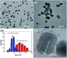
Multimodal imaging based on nanostructures has become a subject of interest for numerous biomedical laboratories. The main focus was placed on applying nanocrystals for the purpose of two types of clinical imaging (contrast and fluorescent agents) due to their excellent luminescence and/or paramagnetic properties. Such systems should also be characterized by low toxicity and high cellular uptake efficiency. Since bare rare earth fluoride nanocrystals influence the cell membrane integrity, it is expected that their coatings will improve biocompatibility profile, as well as increase hydrophilicity, dispersion and chemical stability. Hence, by synthesis of β-NaGdF4:Yb3+Er3+ nanorods (NRs) coated with noncovalently bounded polyethylene glycol monooleate (PEG-Mo), it should be possible to obtain multimodal imaging biomarkers meeting established criteria. Synthesis of β-NaGdF4:Yb3+Er3+@PEG-Mo NRs was performed by the co-precipitation method. These nanostructures were characterized in terms of their size, morphology, zeta potential, magnetic and optical properties as well as their cytotoxicity profile and cellular internalization was evaluated. It was shown that the shape and size of nanocrystals, namely 20 nm nanorods, present generally accepted parameters for biomedical purpose. Ligand attraction of PEG-Mo 860 resulted in the encapsulation of oleic acid coated NRs and formation of hydrophilic bilayer. Superparamagnetic and luminescence properties were highly efficient. Cytotoxic profiles of normal and cancer cell lines were low and determined by dose and time. Cellular uptake was confirmed by the presence of upconversion luminescence in cell interior. These findings are showing multimodal imaging properties of rod shaped β-NaGdF4:Yb3+Er3+@PEG-Mo NRs which may be useful in some biomedical applications.
 Please wait while we load your content...

